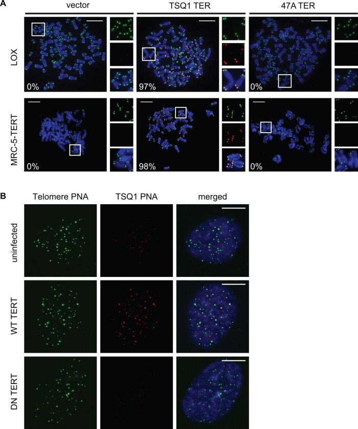
Figure 1.
Robust telomeric incorporation of TSQ1 variant repeats. (A) Metaphase spreads of LOX (top) and MRC-5-TERT (bottom) cells 5 days after infection with control lentivirus (vector) or lentivirus expressing TSQ1 or 47A mutant TER. Wild-type telomeric repeats (green) and TSQ1 variant repeats (red) were detected by FISH. Regions in white boxes are enlarged to the right of the corresponding image. For each condition, between 24 and 35 nuclei were manually scored in a blinded manner and the percentage of TSQ1-positive metaphases is indicated in the bottom left corner. (B) FISH analysis of TSQ1-expressing MRC-5 cells infected with lentivirus expressing wild-type (WT) or catalytically dead dominant-negative (DN) TERT. Cells were fixed 2 days after infection and analysed for colocalization of TSQ1 (red) and wild-type (green) telomeric repeats. At least 30 nuclei from each group were blindly scored, and TSQ1 addition was exclusively observed in cells receiving WT TERT. Scale bars, 10 μm.

