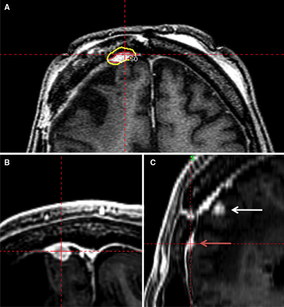
Fig. 3.
a Targeting of atypical meningioma. Axial SPGR MRI sequence with radiosurgical dose targeting enhancing tumor. The yellow line represents the 50% isodose line. The red line represents tumor delineation. b, c Follow-up MRI demonstrating marginal recurrence. b An axial SPGR MRI sequence. c Sagittal SPGR MRI sequence

