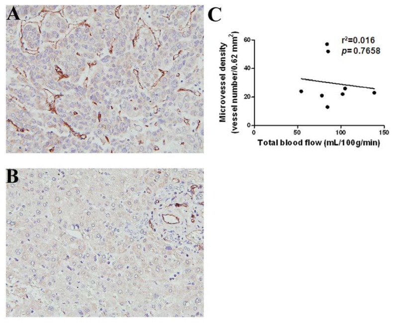
Figure 3.
Histology and immunohistochemistry (IHC) of HCC and surrounding non-tumor liver parenchyma. (A) HCC section showed a higher numbers of CD31-positive endothelial cells (deep brown color) by IHC; (B) The section of the surrounding non-tumor region of liver parenchyma shows low CD31-positive endothelial cells by IHC; (C) Representative CT perfusion parameter (total BF) and average microvessel density (MVD), estimated by counting CD31-positive microvessels, did not show any significant trend. Microvessels were counted on a 200× field (0.62 mm2).

