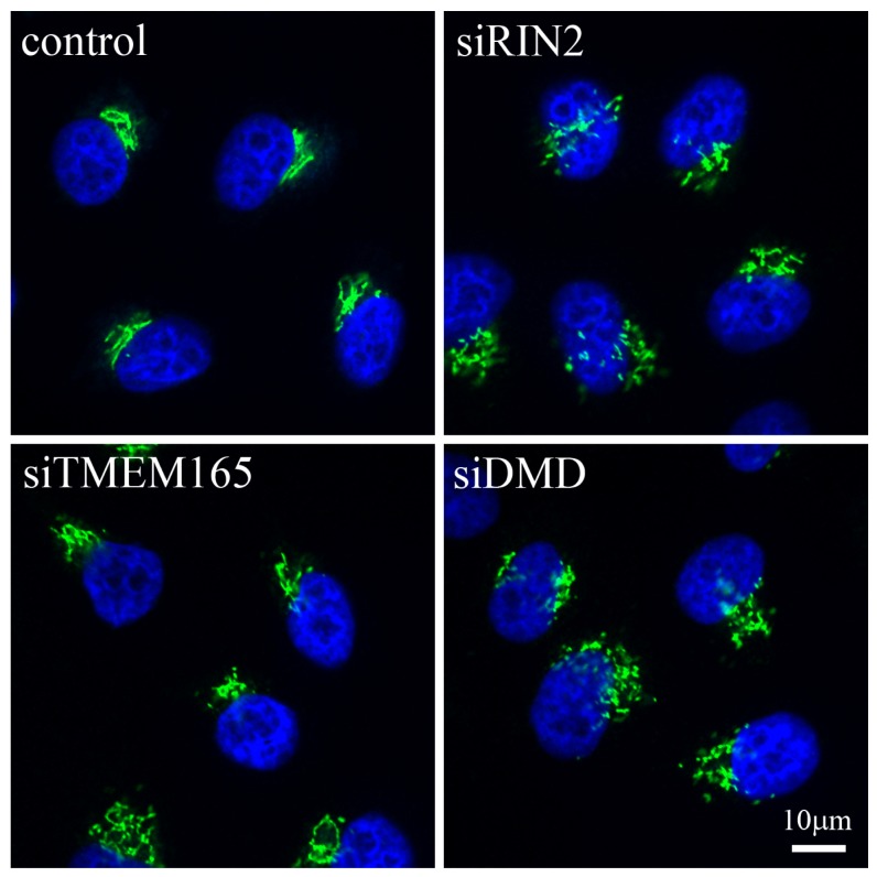
Figure 1.
Immunofluorescence showing gross changes in Golgi complex morphology in response to the down-regulation of particular proteins. HeLa cells were incubated with RNAi reagents targeting RIN2, TMEM165 or DMD and incubated for 48 h. Cells were fixed and stained for the Golgi matrix protein GM130 (green) and the nucleus (blue). A normal compact juxta-nuclear Golgi complex can be seen in control cells, whereas the cells depleted for the three proteins of interest show varying degrees of change in Golgi complex morphology. Scale bar corresponds to 10 μm.

