
Figure 7.
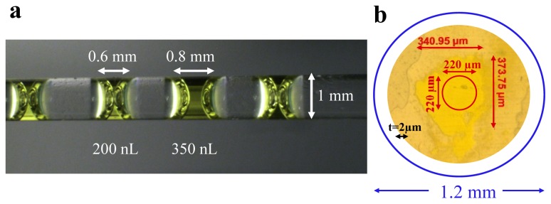
Protein samples prepared for time-resolved spectroscopic experiments. (a) Capillary containing multiple drops of PYP solution; four 200–400 nL drops of protein solution are mounted together; (b) PYP crystal crushed between two cover slides. The relative sizes of the laser pulse (blue) and the Xenon light source (red) are shown. The sample has a thickness (t) of approximately 2 μm.