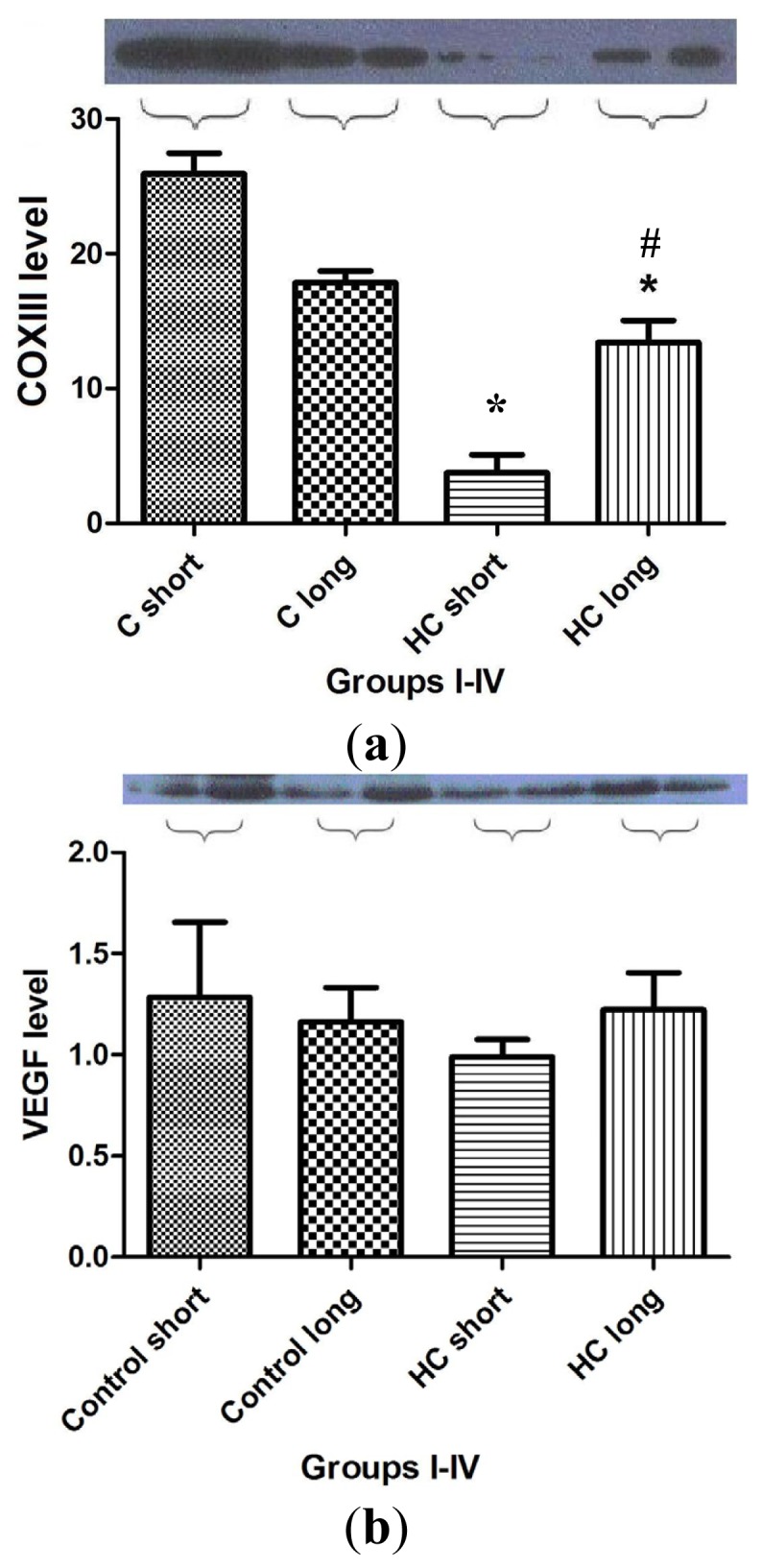
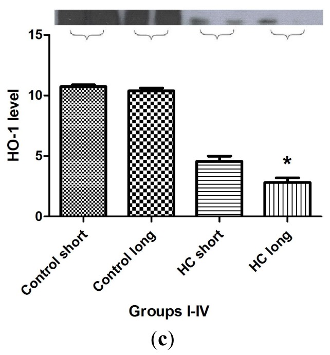
Figure 7.

Western blot analysis for biomarkers of cardiac tissue function. Expression of COX III (7a), VEGF (7b) and HO-1 (7c) protein in left ventricular rabbit myocardium was measured in homogenized cardiac tissue samples drawn from hearts harvested from four groups of rabbits (n = 4 per group), administered feed containing normal chow for 12 weeks (control short); normal chow for 40 weeks (control long); 2% cholesterol during a 12-week time-course (HC short); or 2% cholesterol during a 40-week time-course (HC long). GAPDH expression level was measured as a reference protein. Western blot analyses were conducted on each tissue homogenate in triplicate, and the signal intensity of the resulting bands corresponding to proteins of interest was measured using the Scion for Windows Densitometry Image program, version Alpha 4.0.3.2 (Scion Corporation, MD, USA). Tissue content of each protein is shown in arbitrary units as the mean for each group of rabbits ± SEM. *p < 0.05 for comparison of average levels of COX III and HO-1 in myocardium of HC long animals with all other groups. # p < 0.05 for comparison of average levels of COX III in myocardium of HC long animals with HC short animals. (a) COXIII protein expression in rabbit myocardium; (b) VEGF protein expression in rabbit myocardium; and (c) heme oxygenase-1 (HO-1) protein expression in rabbit myocardium.

