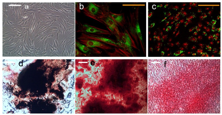
Figure 2.
Morphology of undifferentiated hMSCs (a); magnetic nanoparticle (MNP) targeting PDGFRα (b) and integrin ανβ3 (c) in hMSCs, and histology analyses of hMSCs after 21 days of osteogenic induction (b–d). (a) Light microscope image, scale bar = 200 μm; (b) Confocal image of dextran (green) and F-actin (red) staining showing PDGFRα antibody functionalized MNP targeting in hMSCs, scale bar = 100 μm; (c) Confocal image of dextran (green) and nucleus (red) staining showing RGD peptide functionalized MNP targeting in hMSCs; (d) Alkaline phosphatase and von Kossa staining; (e) Alizarin red s staining; (f) Sirius-red staining. (c–f) Scale bar = 200 μm.

