Figure 1.

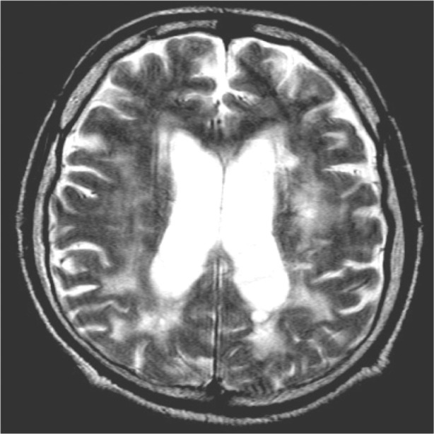
Magnetic resonance imaging scan of the brain of the first patient (TQ, 44 years old; axial section) showing diffuse hyperintensities involving the white matter, mainly in the posterior cerebral areas.
Abbreviation: TQ, first patient.

Magnetic resonance imaging scan of the brain of the first patient (TQ, 44 years old; axial section) showing diffuse hyperintensities involving the white matter, mainly in the posterior cerebral areas.
Abbreviation: TQ, first patient.