
Figure 1.
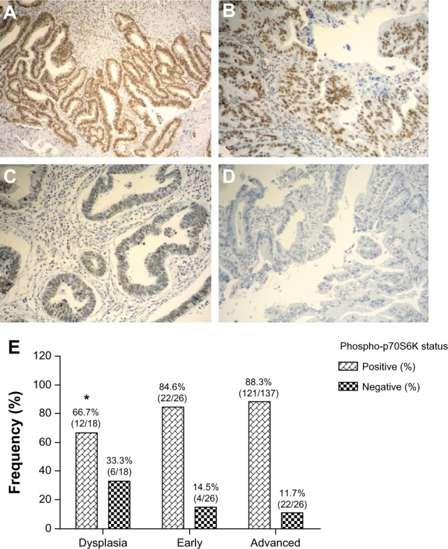
Immunohistochemical staining of phospho-p70S6K in gallbladder tissue. Representative images from gallbladder lesions showing intense (A), moderate (B), weak (C), and negative intensity (D) staining of phospho-p70S6K. Images represent original magnification 100×. (E) Frequency distribution for positive staining of phospho-p70SS6K in sequential gallbladder lesions.
Note: *Compared with advanced gallbladder cancer (P < 0.05).