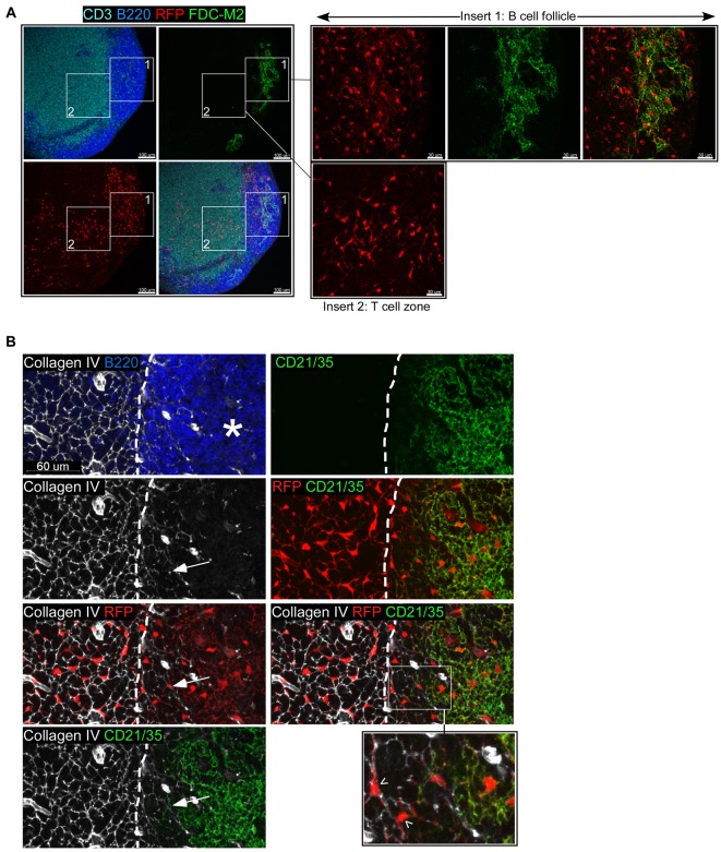
Figure 1. Visualization of a new subset of LN stromal cells.
Confocal images of LN sections from a CD21cre-RFP chimera stained for (A) FDC-M2 (green), CD3 (light blue), and B220 (dark blue) and (B) CD21/35 (green), Collagen IV (white), and B220 (blue). RFP+ cells appear in red. In (B), the star (*) signals the position of the collagen-poor central region of the follicle populated by the CD21+ RFP+ FDC network; arrows indicate the extensions of the conduit network within the follicle and arrowheads point to CD21− RFP+ cells attached to these conduits. The dashed line represents the delineation of the B220 staining. No RFP signal was detected in CD21cre−RFP+ chimeric mice, ruling out a leaky expression of the reporter. Data are representative of three different experiments (two mice per experiment).

