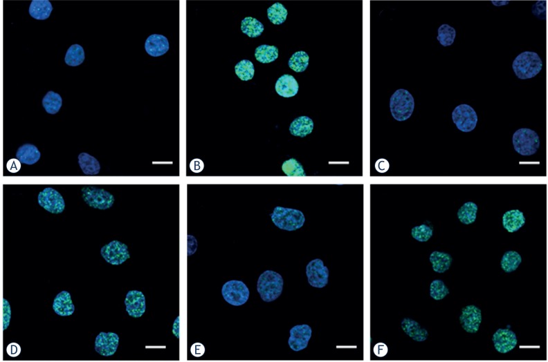
FIGURE 4.
Digitized images of γH2AX foci in Hela, HepG2 and MEC-1 cell lines. After exposure to 4 Gy 12C6+ followed incubated for 30 min, cells were grown and irradiated on cover slips. DNA was stained with DAPI and γH2AX was detected using an Alexa488- conjugated secondary antibody after staining using a monoclonal anti-γH2AX antibody. (A. Hela-Control; B. Hela-4Gy; C. HepG2-Control; D. HepG2-4Gy; E. MEC-1- Control; F. MEC-1-4Gy. Scale bar, 15um)

