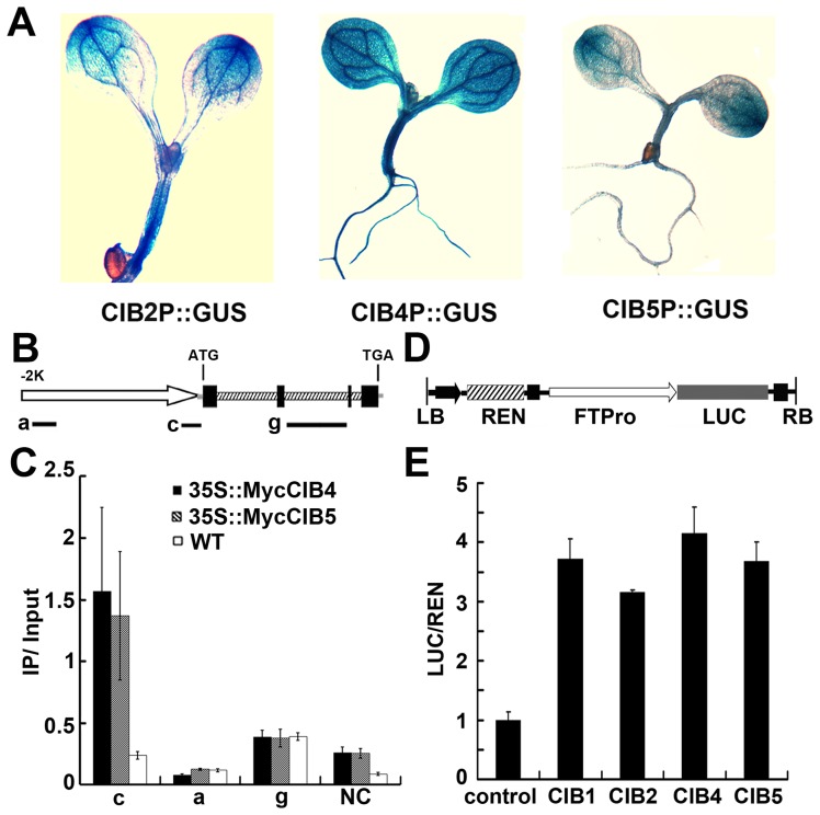
Figure 4. ChIP-qPCR showing interaction of CIB4 and CIB5 with chromatin regions of the FT gene.
(A) GUS staining of seedlings expressing CIB2::GUS, CIB4::GUS, CIB5::GUS transgene. (B) A diagram depicting the putative promoter (arrow), 5′ UTR (grey line), exons (black boxes), introns (dashed boxes), 3′ UTR (grey line) of the FT gene. Black solid lines depict the DNA regions that were amplified by ChIP-PCR using the indicated primer sets. (C) Representative results of the ChIP-qPCR assays. Chromatin fragments (∼500 bp) were prepared from 7-day-old transgenic seedlings expressing 35S::Myc-CIB4 or 35S::Myc-CIB5, immunoprecipitated by the anti-Myc antibody, and the precipitated DNA were qPCR-analysised using the primer pairs indicated. The IP/input ratios are shown with the standard deviations (n≥3). (D) Structure of the FT promoter–driven dual-Luc reporter gene. 35S promoter (black arrow), FT promoter (−2000 bp–0 bp) (white arrow head), REN luciferase (REN), firefly luciferase (LUC), and T-DNA (LB and RB) are indicated. (E) Relative reporter activity (LUC/REN) in planta with different effectors (CIB1/2/4/5) expression. Control: transiently expressed reporter only, CIB1: transiently expressed reporter and CIB1, CIB2: reporter and CIB2, CIB4: reporter and CIB4, CIB5: reporter and CIB5. Tobacco leaves were transfected with the reporter and the effector (CIB1 or CIB2 or CIB4 or CIB5); kept in white light for 3 days. The relative LUC activities normalized to the REN activity are shown (LUC/REN, n = 3).

