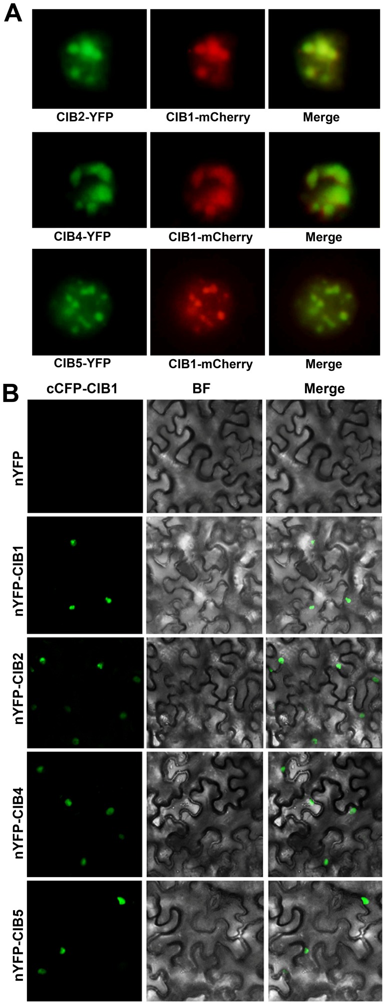
Figure 5. CIB1 interacts with CIBs.
(A) Fluorescent microscopy images showing that CIB2, CIB4 and CIB5 (Green) all co-localize with CIB1 (Red) in the nucleus. (B) Bimolecular fluorescence complementation assays of the in vivo protein interaction. Leaf epidermal cells of N. benthamiana were cotransformated with cCFP–CIB1 and nYFP [31], or nYFP-CIB1, or nYFP-CIB2, or nYFP-CIB4, or nYFP-CIB5. BF, Bright Field; Merge, overlay of the YFP and bright field images.

