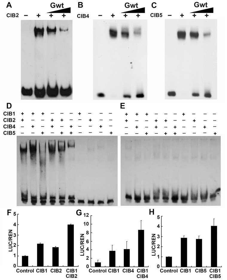
Figure 6. CIB heterodimers bind to the non-canonical E-box sequence of the FT promoter.
(A–C) Competitive electrophoretic mobility shift assay (EMSA) showing binding of CIB2 (A), CIB4 (B), and CIB5 (C) to the G-box DNA (canonical E-box) in vitro. Relative amounts of the un-labeled competitive oligonucleotide containing the G-box sequence used in the reactions are indicated on the top. (D–E) An EMSA experiment showing association of the CIB heterodimers, but not monomers, with the non-canonical E-box DNA of the FT promoter (region c in Figure 4B). The indicated CIB proteins were expressed and purified from E. coli, and incubated with the labeled oligonucleotide containing the E-box DNA of the FT promoter (D) or the same sequence except that the E box was replaced with AAAAAA sequence (E) (F–H) Transient assays show CIBs (CIB1/2/4/5) activation of the FTpro::LUC reporter gene. (F) Control: transiently expressed reporter only, CIB1: reporter and CIB1, CIB2: reporter and CIB2, CIB1 CIB2: reporter, CIB1 and CIB2 together. (G) CIB4: reporter and CIB4, CIB1 CIB4: reporter, CIB1 and CIB4. (H) CIB5: reporter and CIB5, CIB1CIB5: reporter, CIB1 and CIB5. Tobacco leaves were transfected with the reporter and the effectors; kept in white light for 3 days. The relative LUC activities normalized to the REN activity are shown (LUC/REN, n = 3). Error bars indicate SD of three biological repeats.

