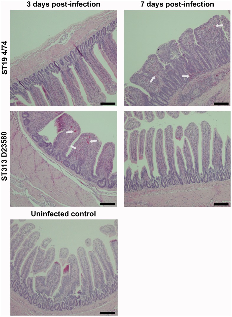
Figure 3. Histopathological changes in the ileum following infection with S. Typhimurium ST313 or ST19.
Photomicrographs (×400 magnification) of histological changes in H and E stained fixed ileal tissue following infection with S. Typhimurium ST313 show a rapid inflammation at three days post-infection leading to villus fusion and flattening accompanied by infiltration of lymphocytes and polymorphonucelar cells (heterophils) and crypt hyperplasia. By seven days post-infection, inflammation and pathology is reduced. In birds infected with ST19 (4/74) S. Typhimurium the inflammatory process appears slower with limited inflammation at three days post infection, but substantial damage to the structure of the ileum at seven days post-infection with a high degree of villus damage and large areas of inflammatory infiltration. Arrows indicate heterophil influxes. Scale bar = 100 µm.

