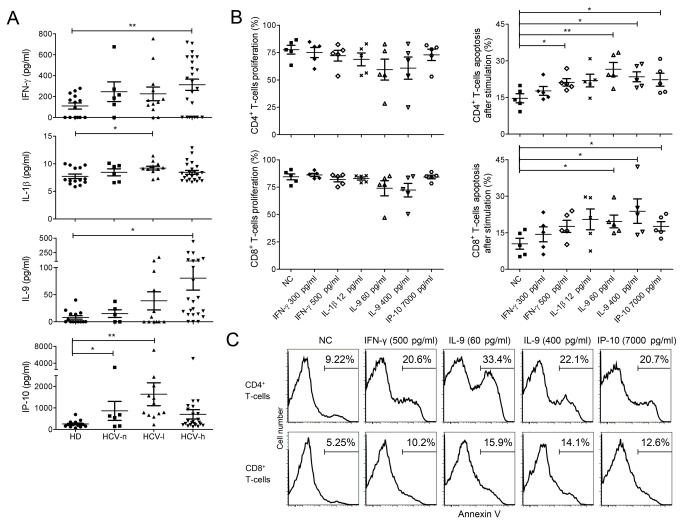
Figure 4. Elevated cytokine levels in the plasma of CHC patients and their roles in sensitizing T-cells to activation-induced apoptosis.
(A), Cytokine levels, including IFN-γ, IL-1β, IL-9, and IP-10, in the plasma of CHC patients and HDs as measured by Luminex assay. (B), Effect of pre-incubation of PBMCs with the indicated cytokines on T-cells proliferation as measured by CFSE dilution and T-cells apoptosis as measured by Annexin-V staining. PBMCs from HDs were pre-incubated with or without (NC) the indicated cytokines for 48 hrs and then stimulated with anti-CD3/CD28 for 3 days before the measurement of proliferation and apoptosis. Data were from 5 independent HDs. (C), Representative FACS profiles of CD4+ and CD8+ T-cells apoptosis after pre-incubation with cytokines followed by stimulation. *, p<0.05; **, p<0.01.

