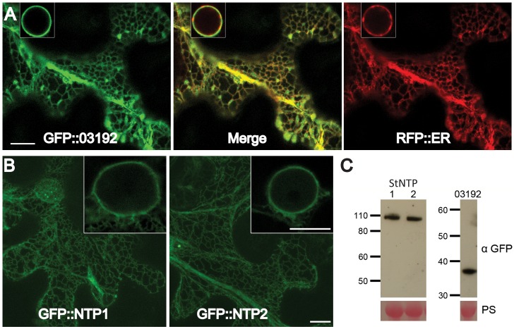
Figure 3. StNTP1, StNTP2 and Pi03192 are localised to the ER membrane in planta.
(A) GFP-Pi03192 co-localises to the ER membrane with an RFP tagged ER marker. Scale bars indicate 10 µm, and insert-images show ER around the nucleus. (B) GFP-StNTP1 and 2 localise to the ER and are found in this membrane surrounding the nucleus (insert-images). Scale bars indicate 10 µm. (C) Immunoblots of GFP-StNTP1, GFP-StNTP2 and GFP-Pi03192 showing the stability of the full length constructs when probed with a specific GFP antibody. PS is Ponceau stain.

