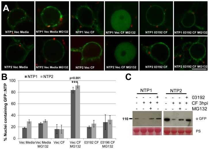
Figure 8. Pi03192 prevents CF-mediated accumulation of StNTP1 and StNTP2 in the nucleus.
(A) Confocal images of localisation of either GFP-StNTP1 or GFP-StNTP2 co-expressing either pFlub (vec) or pFlub-03192 and treated with Media or culture filtrate, plus and minus MG132. Scale bar is 10 µm. (B) Graph shows the percentage of nuclei containing either GFP-StNTP1 or GFP-StNTP2 fluorescence with each treatment. Error bars are standard error and one-way ANOVA analysis shows that treatment with CF and MG132, when co-expressed with empty vector (vec), is statistically different from the other treatments, which are not different from each other. Significance is indicated by p-value and asterisks. (C) Immunoblots show the stability of GFP-StNTP1 and GFP-StNTP2 with different treatments as indicated, probed with a specific GFP antibody. PS is Ponceau stain.

