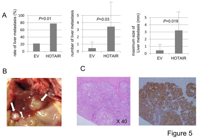
Figure 5. The association of HOTAIR expression with liver metastasis.
A, The effect of HOTAIR on metastasis was investigated by tail vein assay. 1.0 x 10⁵ of HOTAIR expressing cells (MKN-HOTAIR, n=9) and control cells (MKN-EV, n=9) were injected into the tail vein of mice. MKN-HOTAIR cells more frequently showed liver metastases (P=0.01) compared to MKN-EV cells (left panel). In addition, the numbers (middle panel) and sizes of the metastatic liver tumors (right panel) were significant larger with MKN-HOTAIR cells than with MKN-EV cells (P=0.03 and P=0.019, respectively). B, The metastatic tumors could be clearly recognized macroscopically (arrow). C, The tumor cells formed glands and were thought to be derived from the injected human gastric cancer cell lines (left panel, HE, orginal magnification x40). These tumor cells showed intense immunoreactivity for human cytokeratin (right panel).

