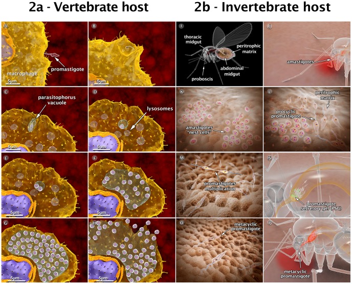
Figure 2. Schematic 3D view of the phases of interaction between the Leishmania amazonensis parasite and vertebrate cells (2a), and between the parasite and the sandfly (2b).
(A) Attachment of a promastigote to the macrophage surface. (B) The process of internalization via phagocytosis begins with the formation of pseudopods (C), leading to the formation of the parasitophorous vacuole (PV). In the PV, the promastigote transforms into an amastigote. (D) Recruitment and fusion of host cell lysosomes with the PV takes place. (E) In the PV, amastigotes divide several times. (F–G) Intense multiplication generates several hundreds of amastigotes. (H) The host cell bursts, and the parasites reach the extracellular space. (I) Schematic view of female sandfly showing the digestive tract. (J) During a blood meal, a female sandfly ingests infected macrophages with amastigote forms present in the blood of the vertebrate host. (K) Amastigotes form “nest cells” in the abdominal midgut. (L) Amastigotes transform into procyclic promastigotes. (M) Promastigotes multiply and attach to the midgut epithelium. (N) Parasites migrate toward the anterior midgut, resume replication and start to produce promastigote secretory gel (PSG). (O) Promastigotes transform into infective metacyclic promastigotes. (P) Metacyclic promastigotes infect a new mammalian host via regurgitation during the blood meal. These images are based on micrographs obtained by scanning and transmission electron microscopy and by video microscopy.

