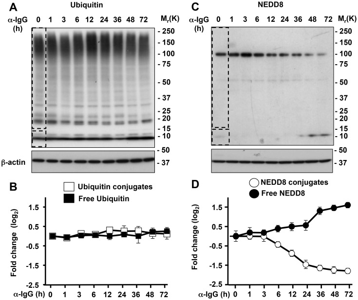
Figure 1. Neddylation is selectively impaired during EBV replication.
Western blots of cell lysates from induced Akata-Bx1 were probed as indicated. A. Western blot illustrating the unchanged levels of ubiquitin conjugates. One representative experiment out of three is shown. B. Quantification of the intensity of the Ub conjugates (dashed rectangle) and free Ub (dashed square) identified in Figure 1A measured by densitometry of the respective areas. The fold change was calculated relative to the intensity at time 0. Mean ± SE of three experiments. C. Western blot illustrating the decrease of NEDD8 conjugates. The prominent band of approximately 100 kD corresponds to neddylated cullins. D. Quantification of the intensity of the NEDD8 conjugates and free NEDD8. Mean ± SE of three experiments.

