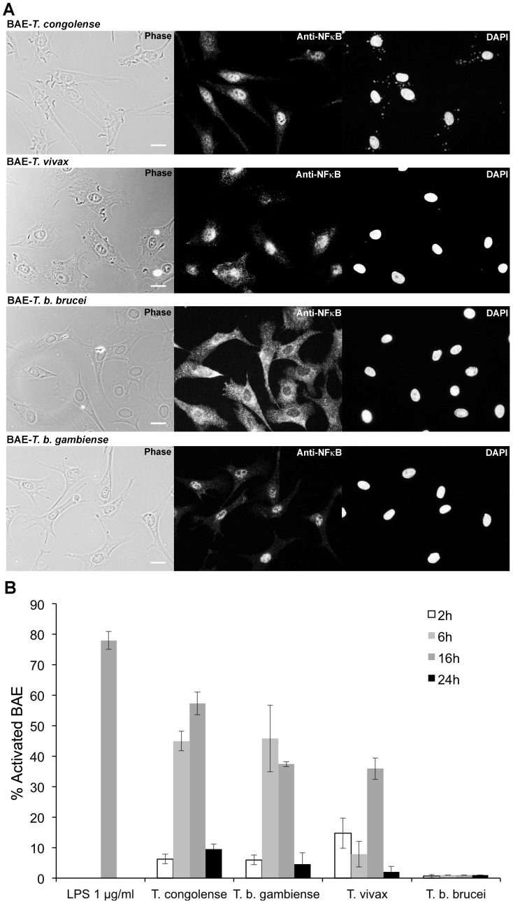
Figure 1. Activation of BAE by African trypanosomes.
(A) NF-κB immunofluorescent staining on BAE after 16 h of coculture with T. congolense IL3000, T. vivax Y486, T. b. brucei AnTat 1.1 and T. b. gambiense 1135 BSF. Scale bars = 20 µM. (B) Kinetics of BAE activation: percentage of activated BAE in presence of 1 µg/ml LPS and after 2, 6, 16 and 24 h of coculture with BSF of T. congolense IL3000, T. vivax Y486, T. b. brucei AnTat 1.1 and T. b. gambiense 1135. Results were similar with T. congolense STIB910 strain, T. b. gambiense LiTat strain, and T. b. brucei 427 strain. Each experiment was repeated at least three times independently. Results are expressed as mean-values±standard deviation (SD).

