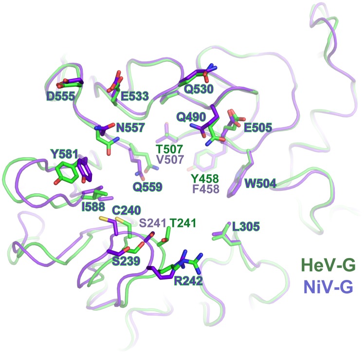
Figure 3. Comparison of the m102.3-binding regions of HeV-G and NiV-G.
The HeV-G (green) and NiV-G (purple) structures are superimposed and viewed from top. The m102.3 contacting residues of HeV-G and the corresponding residues of NiV-G are shown and labeled. Most residues are conserved between HeV-G and NiV-G except for three: T/S241, T/V507 and Y/F458.

