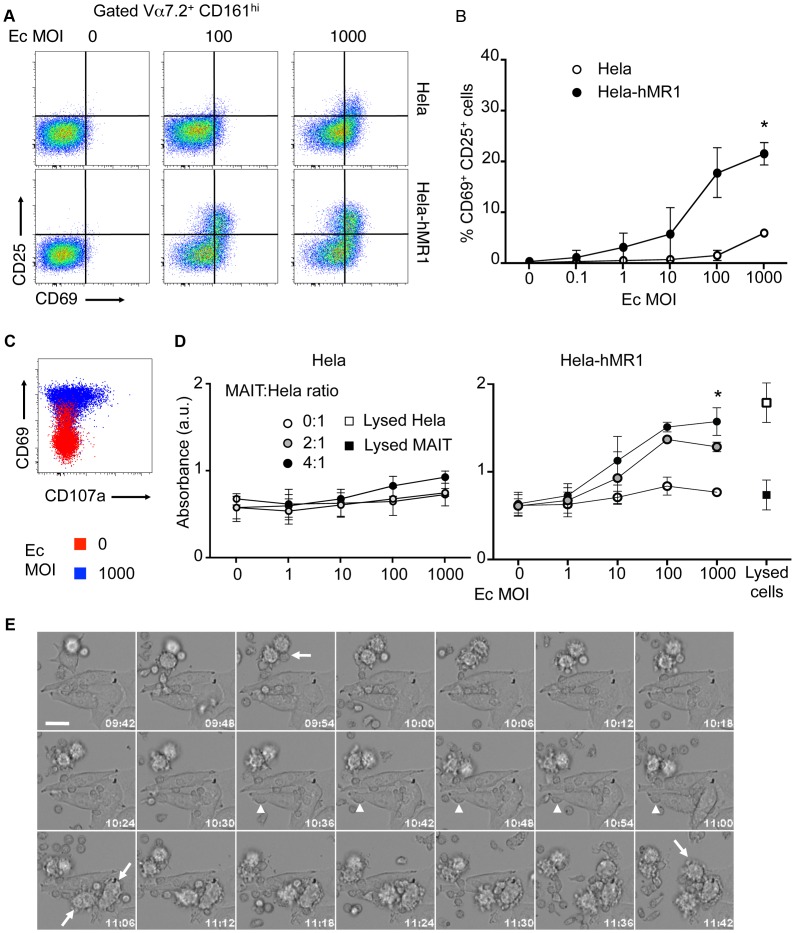
Figure 2. Human MAIT cells are cytotoxic.
(A–B) Activation of purified MAIT (Vα7.2+CD161hi) cells cultured with Hela cells or Hela cells overexpressing the human MR1 protein (Hela-hMR1), in presence of increasing multiplicity of infections (MOI) of Escherichia coli (Ec). (A) FACS analysis of CD69 and CD25 upregulation. (B) Data are mean and SEM of three independent experiments as in A. * indicates statistical significance for the 2 parameters (MOI dose-response curve and Hela vs Hela-MR1) by 2 way ANOVA. (C) Degranulation assessed by CD107a (LAMP1) staining at the cell surface (red: uninfected control; blue: MOI of 1000). (D) Cytotoxicity of MAIT cells cultured with Hela or Hela-hMR1 cells as in A, with several effector∶target ratios (MAIT∶Hela), assessed by quantification of LDH release in the supernatants after overnight coculture. Maximum LDH release after chemical-induced lysis from Hela-hMR1 cells or MAIT cells is represented (white and black squares, respectively). Data are mean and SEM of two independent experiments. * indicates statistical significance for the 2 parameters (MOI dose-response curve and presence of MAIT or not) by 2 way ANOVA. (E) MAIT cells direct cytotoxicity was visualized by time-lapse analysis of Hela-hMR1 cells cultured in presence of Ec bacterial lysate and sorted MAIT (Vα7.2+CD161hi) cells. Arrows indicate cells entering apoptosis. Arrowheads indicate MAIT cells in prolonged interactions with a target cell. Time indicated on each frame (h∶m). Scale bar: 20 µm.

