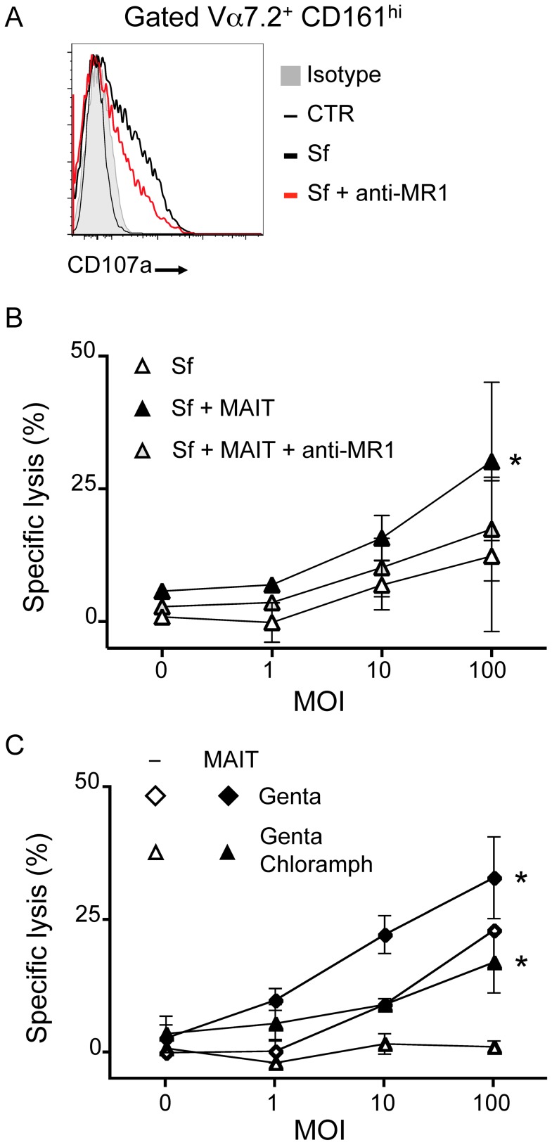
Figure 6. MAIT cells lyse Shigella flexneri-infected epithelial cells.
(A) MAIT cells cultured with Sf-infected Hela cells (as in 5B) degranulate as observed by presence of CD107a at their cell surface (grey: isotype control; thin black line: uninfected; bold black line: Sf MOI 100; red line: Sf MOI 100 with anti-MR1). Representative of two independent experiments. (B) MAIT cells are cytotoxic to Sf-infected Hela cells. Sf-infected Hela cells were cocultured with MACS sorted Vα7.2+ cells and LDH release assessed after overnight culture. LDH release was increased in presence of MAIT cells as compared with Sf-infected Hela alone. This increase is blocked by addition of the anti–MR1 antibody (10 µg/ml). Data are mean and SEM of two independent experiments. * indicates statistical significance for the MOI dose-response curve by 2 way ANOVA. (C) MAIT cell-dependent cytotoxicity is additional to the one induced by the pathogenic Sf. Hela cells infected by Sf were put in medium supplemented with gentamicin (Genta) as in 5B or with both gentamicin and chloramphenicol (Genta Chloramph) before coculture with MACS sorted Vα7.2+ cells. LDH release from target cells was assessed after overnight culture. Data are mean and SEM of two independent experiments.

