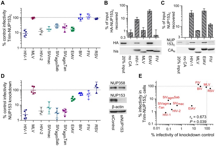
Figure 3. Diverse lentiviruses bind NUP153C.
(A) Transduction efficiencies of retroviral GFP reporter viruses in Trim-NUP153C expressing cells normalized to infection in mock transduced cells, which were set to 100%. Results are the geometric mean of 4 experiments, with error bars denoting 95% confidence intervals. (B) HA-NUP153C expressed in 293T cells was pulled down by the indicated his-tagged retroviral CAN proteins. Captured proteins resolved by SDS-PAGE were western blotted with antibody 3F10 alongside a standard curve of input protein. The results are an average of 5 experiments, with error bars denoting 95% confidence intervals. A representative western blot is shown. (C) SYPRO Ruby detection of retroviral CAN pull-down of purified NUP153C. The results are an average of two experiments, with error bars denoting standard error; one representative gel is shown. (D) (left) Retroviral infectivities in HOS cells knocked down for NUP153 expression as compared to cells treated with a non-targeting short interfering (si) RNA control [19]. The results are the geometric mean of at least 4 experiments, with error bars denoting 95% confidence intervals. (right) Western blot detection of control or NUP153 knockdown HOS cells with antibody mab414, which also detects NUP358. (E) Scatter plot comparing relative retroviral infectivities under each condition.

