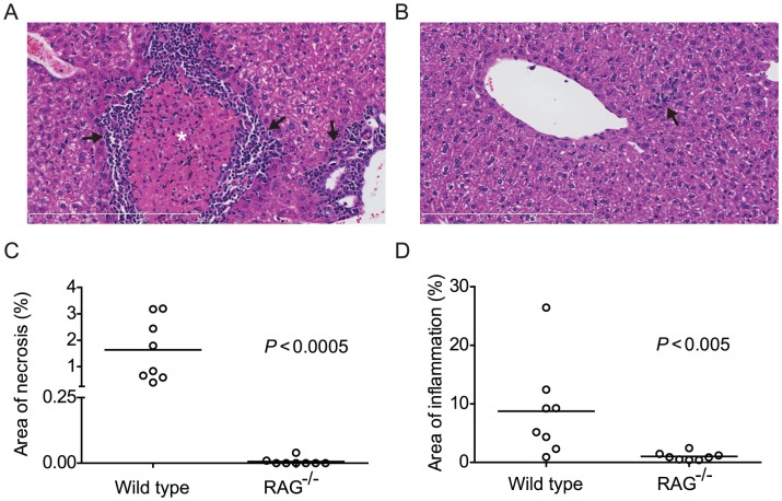
Figure 1. Pre-patent S. mansoni infection does not induce liver necrosis and inflammation in RAG−/− mice.
Wild type (C57BL/6) and RAG−/− mice were infected with S. mansoni cercariae via percutaneous tail exposure and livers were removed for histological analysis at 4 weeks p.i. (A) Representative H&E-stained liver section from a wild type mouse, exhibiting inflammatory cell infiltration (arrows) in periportal area and surrounding a focus of coagulative necrosis (⋆). (B) Representative H&E-stained liver section from a RAG−/− mouse. Note small and isolated cluster of inflammatory cells (arrow). (C) Average percent area of tissue section occupied by coagulative necrosis was calculated for each animal (n = 8, pooled from two independent experiments). Horizontal bars represent mean values for each experimental group. (D) Average percent area of tissue section occupied by inflammatory infiltrate was calculated for each animal (n = 8, pooled from two independent experiments). Horizontal bars represent mean values for each experimental group. P values were determined using the Mann Whitney test. Scale bars in A and B are 300 µm in length.

