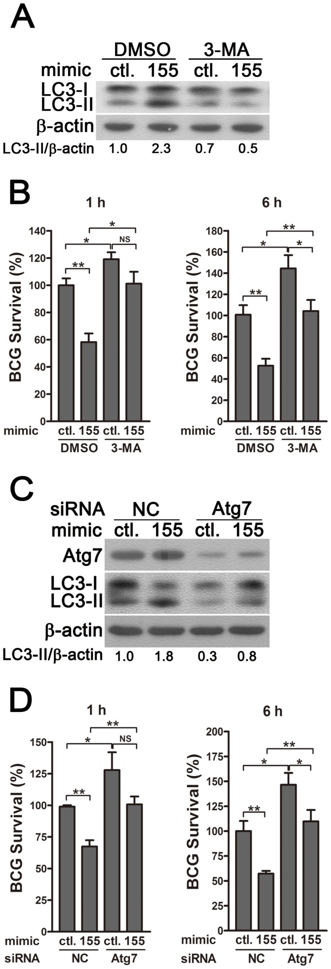
Figure 7. miR-155-induced autophagy promotes the elimination of intracellular mycobacteria.
(A and B) RAW 264.7 cells were transiently transfected with control or miR-155 mimic for 24 h, and then incubated with DMSO or 3-MA for 2 h. Protein levels of LC3 were detected by Western-blot in uninfected cells (A). Intracellular mycobacterial viability was determined at the indicated timepoints by CFU assay after BCG challenge for 1 h (B). (C and D) RAW 264.7 cells were transiently co-transfected with control or miR-155 mimic together with a control siRNA or Atg7 siRNA. The expression levels of Atg7 and LC3 were detected by Western-blot (C). Intracellular mycobacterial viability was determined by CFU assay at the indicated time after challenging with BCG for 1 h (D). Values of LC3-II/β-actin ratios are indicated below the representative blot. Data are shown as the mean ± SEM of three independent experiments. *, p<0.05; **, p<0.01; NS, not significant.

