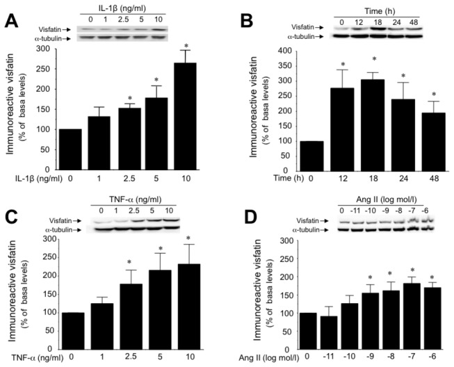
Figure 1. Inflammation enhances intracellular visfatin levels in HUVEC.

(A) Concentration-dependent effect of IL-1β (1 to 10 ng/mL; 18 h) on cellular visfatin content determined by Western blotting. (B) Time course of visfatin induction by IL-1β (10 ng/mL) over 48 h. Visfatin content was also determined in HUVEC challenged for 18 h with either (C) TNF-α (1 to 10 ng/mL) or (D) Ang II (10 pmol/L to 1 µmol/L). Data are the mean±SEM of five independent experiments. *P<0.05 vs non-stimulated cells. Representative gels are shown on the top.
