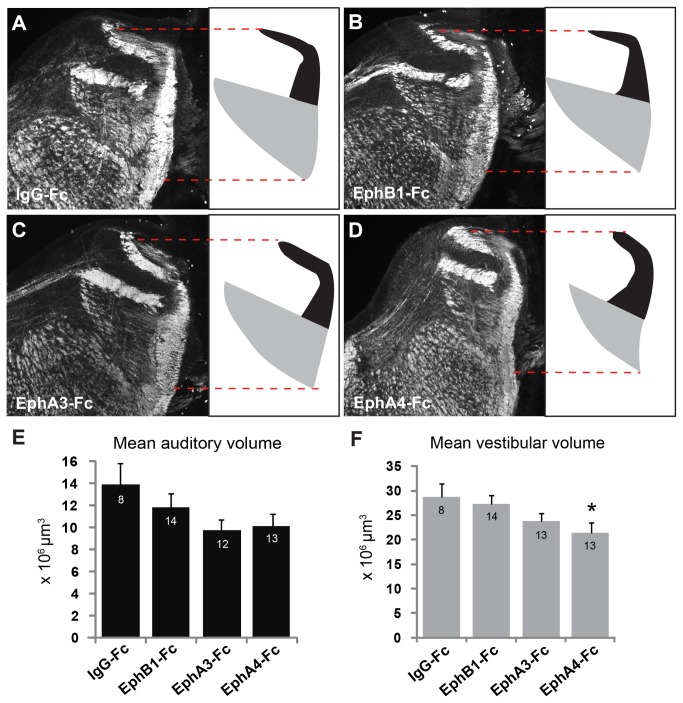
Figure 3. Eph receptor inhibition causes reduction in hindbrain compartment.
(A-D) Neurofilament immunofluorescence (left) of hindbrain sections at level of VIIIth nerve entry with auditory (black) and vestibular (gray) components schematized (right). Red dashed line indicates entire dorsoventral extent of the VIIIth nerve projections in hindbrain. Volumes found in IgG-Fc (A) EphB1-Fc (B), and EphA3-Fc (C) did not significantly differ. (D) Vestibular hindbrain volume is reduced following EphA4-Fc treatment. (E,F) Graphs of mean auditory (E) and vestibular (F) volumes for all treatment groups. Numbers of samples are indicated in each bar; asterisk indicates p <0.05.

