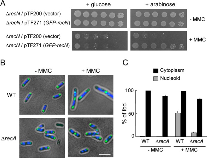
FIGURE 1.

RecN foci in wild-type and ΔrecA cells with or without DNA damage. A, ΔrecN cells carrying either an arabinose-inducible GFP-recN gene (pTF271) or a pBAD vector (pTF200) were diluted and spotted onto LB plates with or without MMC (0.5 μg/ml) in the presence of either glucose or arabinose. B, the subcellular localization of GFP-RecN foci in response to MMC-induced damage. Wild-type or ΔrecA cells carrying pTF271 were exposed to MMC (0.5 μg/ml) followed by the addition of arabinose (0.05%, w/v) to induce GFP-RecN. The panels show GFP/DAPI-merged images of cells 30 min after the addition of arabinose. Scale bar indicates 2.5 μm. C, quantitative analysis of GFP-RecN foci. For cells incubated with or without MMC, ∼150 cells were examined. The results represent the average of at least three independent measurements. Error bars indicate S.D.
