
FIGURE 4.
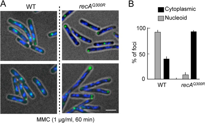
Nucleoid-associated RecN foci in response to MMC-induced DNA damage. A, MMC damage-induced RecN foci in recAQ300R cells. Cells carrying the SOS-inducible GFP-recN (pSG101) were exposed to MMC for 60 min. The panels show GFP/DAPI-merged images of cells. Scale bar indicates 2.5 μm. B, quantitative analysis of GFP-RecN foci. For cells with MMC damage, >150 cells were examined. The results represent the average of at least three independent measurements. Error bars indicate S.D.