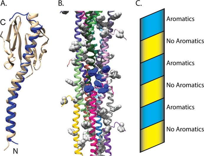
FIGURE 5.

Model of a nanowire fiber based on the structure of G. sulfurreducens PilA. A, superimposition of PilA from G. sulfurreducens on to the homologous type IV pilin from N. gonorrhoeae (Protein Data Bank code 2HIL) (12). The amino terminus and carboxyl terminus are indicated by N and C, respectively. B, model of the bacterial nanowire, based on the pilus assembly of N. gonorrhoeae (Protein Data Bank code 2HIL) (12). Aromatic side chains are shown in space filling. A single cluster of aromatic side chains is shown in blue space filling, whereas all others are shown in gray. Ribbons of each subunit were colored individually. C, schematic diagram showing the progression of the aromatic clusters up the pilus structure. The aromatic band is colored blue, and the aromatic devoid band is colored yellow.
