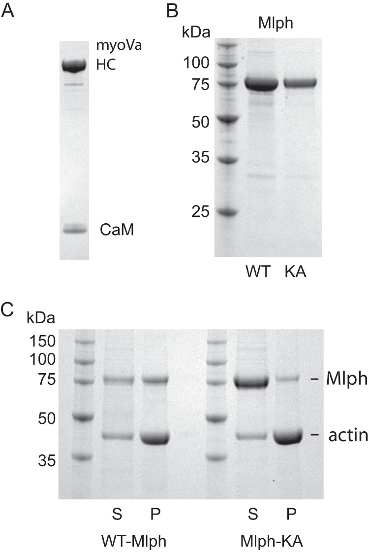
FIGURE 2.

SDS-gels of purified proteins and ability of Mlph to bind actin. A, purified full-length myoVa with an N-terminal biotin tag showing the myoVa heavy chain (HC) and calmodulin (CaM); 4–12% gradient SDS-gel. B, left lane, marker proteins with indicated molecular masses. Middle lane, WT-Mlph. Right lane, mutant Mlph-KA, 12% SDS-gel. C, co-sedimentation of actin with WT-Mlph or Mlph-KA showing that Mlph-KA has a decreased ability to interact with actin. S, supernatant; P, pellet; 12% SDS-gel.
