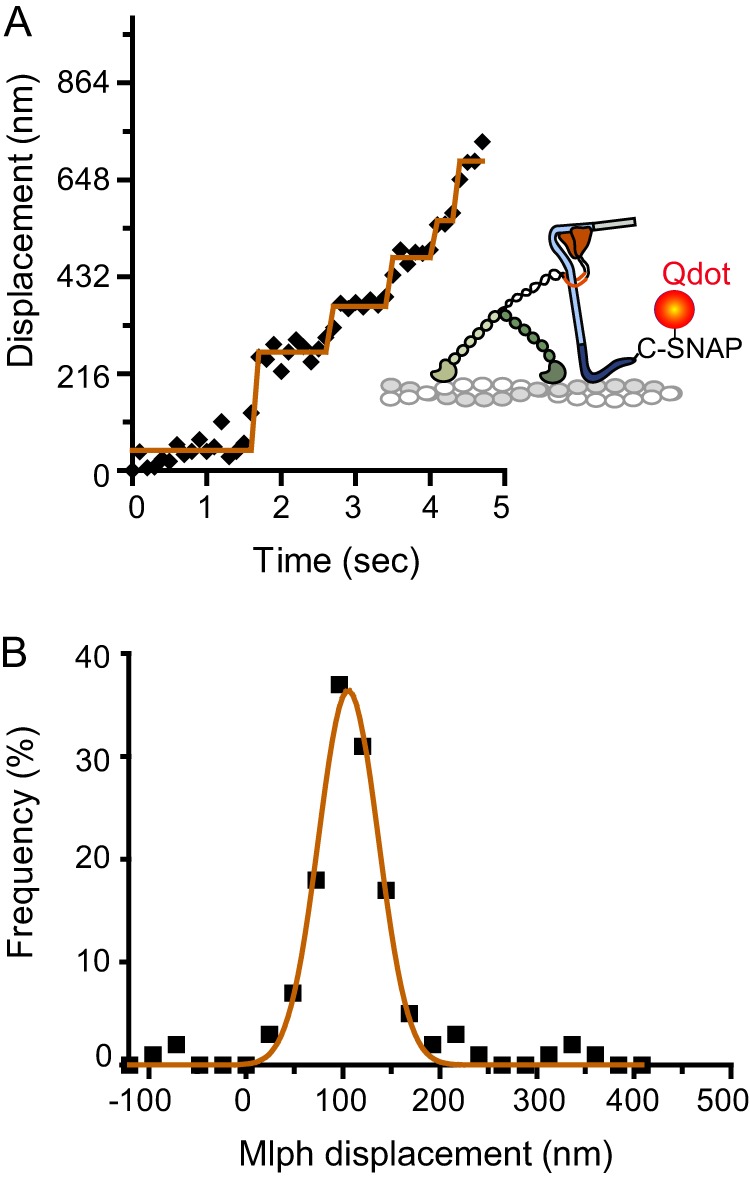
FIGURE 7.

Step-like movement of a Qdot bound to the C terminus of Mlph, near the actin binding site, in complex with unlabeled myoVa. A, Mlph-C-SNAP-biotin was bound to a streptavidin-Qdot. MyoVa did not contain a biotin tag. A representative displacement versus time trace is shown. B, the histogram of Mlph displacement was fit with a single Gaussian (d = 106 ± 31 nm, n = 131).
