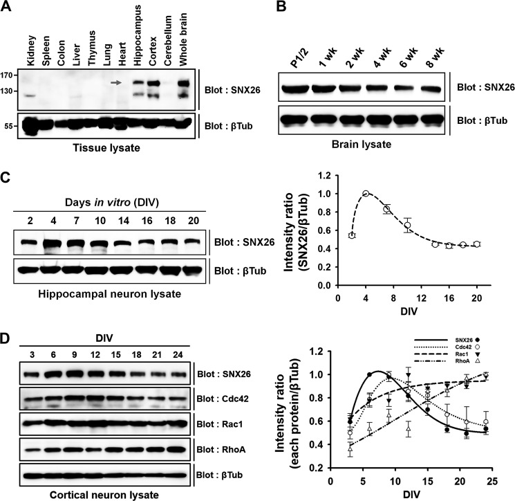
FIGURE 1.
Expression patterns of SNX26 protein in various tissues and during development of postnatal brain and neuron culture. A, tissue distribution of SNX26 protein from 2-week-old mice. Arrow points to the size of SNX26, ∼150 kDa. B, Western blot analysis of postnatal mouse brains. Postnatal brains were lysed, evenly loaded (100 μg) in SDS-polyacrylamide gels, and immunoblotted with a specific anti-SNX26 mouse antibody. SNX26 expression gradually decreases during maturation. C, developmental changes of SNX26 protein expression in cultured rat hippocampal neurons. Cultured primary hippocampal neurons were prepared at the indicated DIV, lysed, and evenly loaded (75 μg) in SDS-polyacrylamide gels. Western blot analysis was performed using a specific anti-SNX26 antibody. SNX26 expression rises during DIV 2–7 and then decreased, and after DIV 16 the levels persisted. D, developmental changes of SNX26, Cdc42, Rac1, and RhoA protein expression in cultured rat cortical neurons. Cultured primary cortical neurons were prepared at the indicated DIV, lysed, and evenly loaded (75 μg) in SDS-polyacrylamide gels. Western blot analysis was performed using each specific antibody. The developmental expression pattern of Cdc42, but not of Rac1 or RhoA, are similar to that of SNX26. βTub, β-tubulin.

