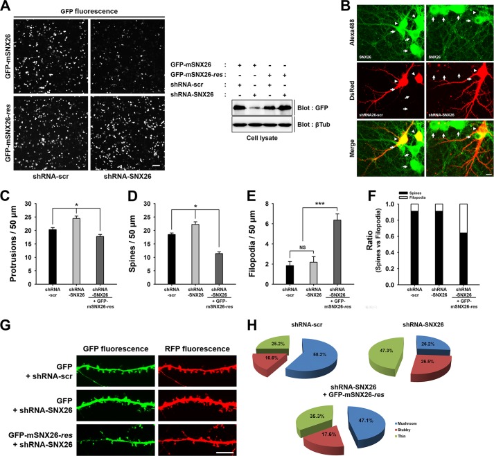
FIGURE 9.
Knockdown of endogenous SNX26 by shRNA causes alterations in dendritic spine formation, which can be partially rescued by resistant mouse SNX26. A, HEK293T cells were co-transfected with GFP-mSNX26 or GFP-mSNX26-res and shRNA-scr or shRNA-SNX26, respectively. The knockdown efficiency of shRNA-SNX26 was confirmed by immunostaining or Western blotting with anti-GFP antibody. Although the expression of GFP-mSNX26 was successfully reduced by shRNA-SNX26, that of GFP-mSNX26-res was not affected by shRNA-SNX26. The term, res, indicates silent mutation that is resistant to shRNA-SNX26. Scale bar, 10 μm; βTub, β-tubulin. B, endogenous expression of SNX26 in cultured hippocampal neurons was reduced by shRNA-SNX26. Hippocampal neurons were transfected with shRNA-scr or shRNA-SNX26 at DIV 8, fixed at DIV 13, and immunostained with anti-SNX26 antibody, followed by Alexa 488-conjugated donkey anti-goat antibody. Alexa 488 channel shows endogenous levels of SNX26. dsRed channel shows the neurons transfected with shRNA-scr or shRNA-SNX26. Arrowheads indicated the soma of neurons transfected with shRNAs. The neuron expressing shRNA-SNX26 showed the reduced levels of SNX26 expression at soma compared with nontransfected neurons (arrows). Scale bar, 10 μm. C–H, cultured hippocampal neurons were transfected at DIV 8 with shRNA-scr or shRNA-SNX26 with GFP or shRNA-SNX26 with GFP-mSNX26-res and fixed at DIV 13. Quantification of the number of total protrusions, spines, and filopodia with expression of each group is shown. Spines were defined as dendritic protrusions of <5 μm in length with heads and filopodia as dendritic protrusions of <10 μm in length without heads. Data were collected from 22 to 42 dendrites of 12–24 neurons for each group in three independent experiments. *, p < 0.01 (ANOVA and Tukey's HDS post hoc test); ***, p < 0.001 (Student's t test). G, representative images of spine morphologies from the dendrite in the neuron expressing shRNA-scr or shRNA-SNX26 with GFP or shRNA-SNX26 with GFP-mSNX26-res, respectively. Scale bar, 15 μm. G, pie graphs illustrating the changes in the portion of different morphological types of spine protrusions in the neurons expressing shRNA-scr or shRNA-SNX26 with GFP or shRNA-SNX26 with GFP-mSNX26-res.

