
FIGURE 5.
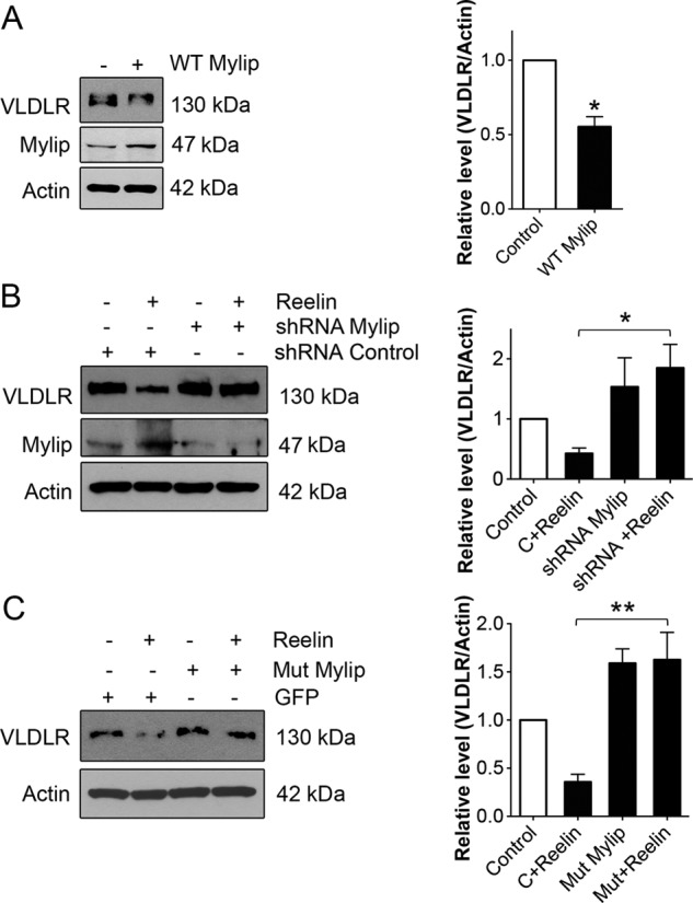
Involvement of Mylip/Idol in the regulation of VLDLR levels by Reelin. Hippocampal neurons were treated as indicated below and further stimulated with 1 μg/ml Reelin for 24 h. VLDLR and Mylip/Idol levels were determined by immunoblotting. β-Actin was used as a control. Left panels, immunoblots; right panels, quantification. A, neurons were infected with adenovirus expressing wild-type Mylip/Idol as described under “Experimental Procedures.” VLDLR levels decreased in the Mylip/Idol-overexpressing cells. Values are means ± S.D. (n = 3). *, p < 0.05 for Reelin versus the control. B, neurons were transfected for 3 days with control (C) scrambled or Mylip/Idol shRNA plasmids for 3 days and further stimulated with 1 μg/ml Reelin for 24 h. Reelin reduced VLDLRs in control shRNA- but not Mylip/Idol shRNA-treated cells. Mylip/Idol levels were down-regulated by >70%. Values are means ± S.D. (n = 3). *, p < 0.05 for the control + Reelin versus shRNA + Reelin. C, neurons were transfected with control EGFP and mutant Mylip/Idol plasmids. Reelin reduced VLDLRs in control but not mutant Mylip/Idol-expressing cells. Mutant Mylip/Idol also increased the basal levels of Mylip/Idol. Values are means ± S.D. (n = 3; ANOVA p value, 0.0023; F, 17.34; Tukey's test). **, p < 0.01 for the mutant + Reelin versus the control + Reelin.