FIGURE 3.
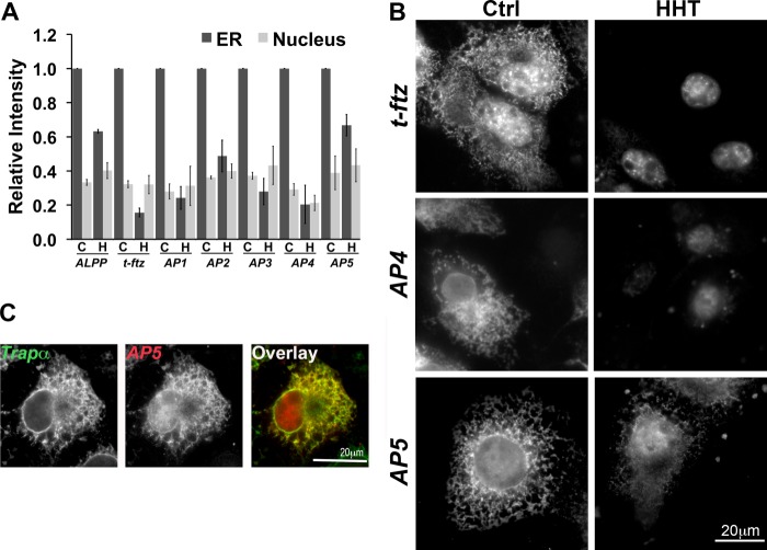
The element responsible for the translation-independent ER localization is present in AP5. COS-7 cells were transfected with plasmid containing either full-length ALPP, t-ftz, AP1, AP2, AP3, AP4, or AP5 and allowed to express mRNA for 18–24 h. The cells were then treated with DMSO (C or Ctrl) or HHT (H) for 30 min and then extracted with digitonin. Cells were then fixed, stained for mRNAs using specific FISH probes (ftz probe was used to detect AP1–5), and imaged. A, quantification of the fluorescence intensities of mRNA in the ER and nucleus. All data were normalized to the ER staining intensity in the control treated group for each construct. The results were normalized to the ER staining intensities in the control treated group for each construct. Each bar represents the average ± S.E. (error bars) of three independent experiments (each experiment consisting of n>30 cells). B, examples of COS-7 cells expressing either t-ftz, AP4, or AP5. C, example of a COS-7 cell expressing AP5 that has been treated with HHT for 30 min then extracted with digitonin. The cell was co-stained for AP5 using ftz FISH probes and Trapα, an ER marker, by immunofluorescence. Scale bars, 20 μm.

