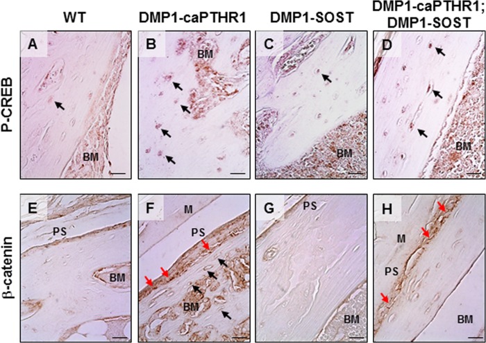
FIGURE 3.
DMP1–8kb-caPTHR1 mice exhibit activation of PTHR/cAMP and PTHR/Wnt signaling in osteocytes. Expression of phosphoSer133-CREB (P-CREB, A–D) and β-catenin (E–H) was detected by immunohistochemistry in sections of tibial bone. Bone surfaces facing the periosteum (Periosteal Surface, PS) and the bone marrow (BM) and muscle fibers (M) are indicated. In some sections the bone is detached from the surrounding muscle or bone marrow tissue. Black arrows point to osteocytes labeled with anti-P-CREB (A–D) or anti-β-catenin (E–H) antibodies. Red arrows point to osteoblasts on the periosteal surface of bone labeled with anti-β-catenin antibody. Note that β-catenin is detected within the cells in the bone section from DMP1–8kb-caPTHR1 mice (F), but only in the cell membrane in the bone section from double transgenic DMP1–8kb-caPTHR1; DMP1-Sost mice (H). Bars indicate 10 μm.

