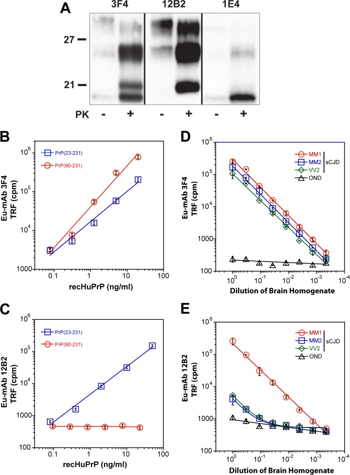
FIGURE 1.
Western blot specificity and calibration of CDI using mAb 3F4 and 12B2. A, WB pattern of typical sCJD with mixed type 1 + 2 PrPSc before and after proteinase K (PK) treatment. The WB was developed with monoclonal antibody 3F4 (epitope 107–112) reacting with the doublet of both 21- and 19-kDa bands of unglycosylated rPrPSc, 12B2 (epitope 89–93) reacting preferentially with 21-kDa band (type 1), and 1E4 (epitope 97–108) reacting preferentially with type 2 rPrPSc. The molecular mass of the standard proteins is in kDa. B and C, calibration of CDI with (blue squares) full-length (PrP(23–231,129M)) or (red circles) truncated (PrP(90–231,129M)) human prion protein and detected with europium-labeled mAb 3F4 (B) or with Europium-labeled mAb 12B2 (C). D and E, sensitivity and specificity of type 1 rPrPSc detection with CDI using europium-labeled mAb 12B2. The different cases of sCJD that were classified pure after diagnostic WBs and a case of other neurological disorders (OND) were serially diluted and tested with CDI using europium-labeled mAb 3F4 (D) or europium-labeled mAb 12B2 (E). To obtain optimum discrimination of type 1 from type 2 rPrPSc, samples were treated with PK at a concentration equivalent to 3 IU/ml (100 μg/ml) of 10% brain homogenate for 1 h at 37 °C and precipitated with phosphotungstic acid after blocking PK with the protease inhibitor mixture. The 8H4 mAb was used for capture of denatured PrPSc. Data points and bars represent average ± S.D. obtained from three or four independent measurements.

