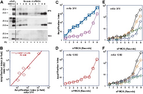
FIGURE 7.

PMCA of mixed type 1 + 2 PrPSc with PrPC(N181Q,N197Q) substrate. The sPMCA was performed with initial 10-fold dilution of type 1 + 2 sCJD brain homogenate followed by limited 2-fold dilution between rounds. A, representative WB was developed with mAb 3F4 for both type 1 and type 2, mAb 12B2 for type 1, and mAb 1E4 for type 2 PrPSc. B, amplification index of total PrPSc and type 1 PrPSc was determined with CDI in six cases of type 1 + 2 sCJD using europium-labeled mAb 3F4 and europium-labeled mAb 12B2, respectively, after 9 rounds of PMCA. C and D, amplification of pure type 1 (red circles) PrPSc and pure type 2 (blue squares) in sPMCA followed by CDI with europium-labeled mAb (C) 3F4 or (D) 12B2 and expressed as an amplification index. E and F, amplification indexes of mixed cases of type 1 + 2 sCJD (diamonds) followed with mAb 3F4 (E) or 12B2 (F).
