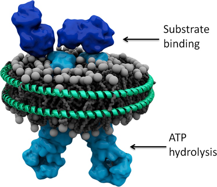
FIGURE 1.

Schematic of OpuA reconstituted in a phospholipid bilayer nanodisc. The schematic is based on the structures of the OpuAC domains (Protein Data Bank codes 3L6G and 3L6H; dark blue (49)) of OpuA, the structure of the membrane domain of the molybdate transporter (Protein Data Bank code 2ONK; light blue (50)), and the structural analysis of the CBS domains of OpuA (15). Arrows indicate the localization of the substrate-binding (extracellular) and nucleotide-binding (cytoplasmic) domains.
