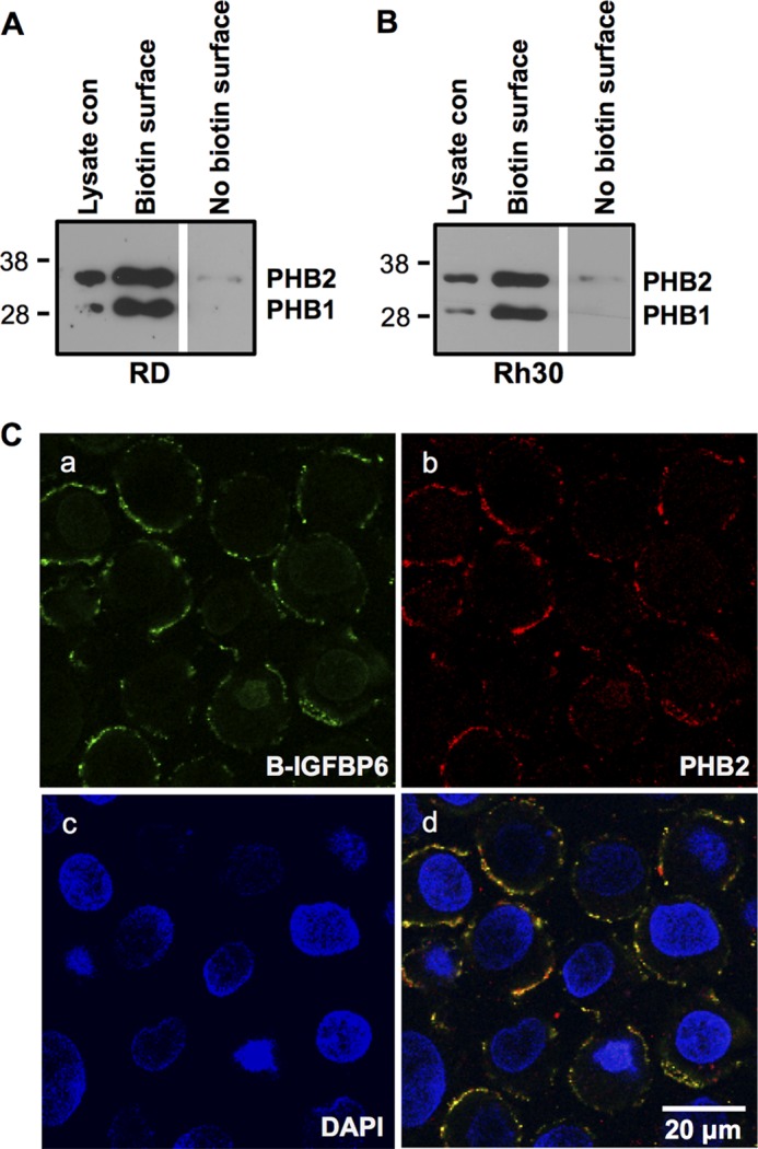
FIGURE 3.

PHB2 and IGFBP-6 colocalize to the plasma membrane in RMS cells. RD (A) or Rh30 (B) cells were subjected to cell surface biotinylation, followed by streptavidin-agarose pull-down and PHB1 and PHB2 Western blotting. Whole lysate control (con) is shown. The blots are representative of three experiments. C, Rh30 cells were incubated with biotinylated IGFBP-6 and PHB2 antiserum, followed by incubation with streptavidin-Alexa Fluro 488 nm and anti-rabbit antibody-Alexa Fluor 567 nm, and then stained with DAPI. Staining is shown for IGFBP-6 (a), PHB2 (b), and DAPI (c). d, merge of a–c.
