Fig. 1.

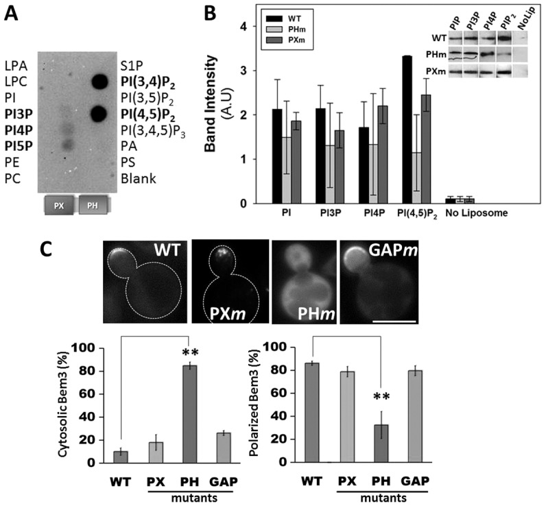
Bem3 occupies cortical membrane sites via interactions with phosphoinositols. Recombinant, purified His6-tagged PX-PH domain fragment of Bem3 was used for binding to PIP microstrips (A) and in liposome floatation assays (B). Detection by western blotting of liposome-bound Bem3 fragments corresponding to a representative experiment is shown in B. (C) Upper panel: cells expressing GFP–Bem3 (WT or mutant) were grown overnight in selective medium at 30°C and imaged at 100× using a FITC filter. Lower panel: localization patterns of GFP-tagged wild-type (WT) and mutant Bem3 were quantified by analyzing at least 200 cells. Data are the total percentage of cells with either a cytosolic (left) or polarized (right) distribution of each construct. Statistical significance was calculated using Student's t-test (**P<0.001). Scale bar: 5 µm. LPA, lysophosphatidic acid; LPC, lysophosphatidylcholine; PA, phosphatidic acid; PC, phosphatidylcholine; PE, phosphatidylethanolamine; PI, phosphatidylinositol; PS, phosphatidylserine; S1P, sphingosine 1-phosphate.
