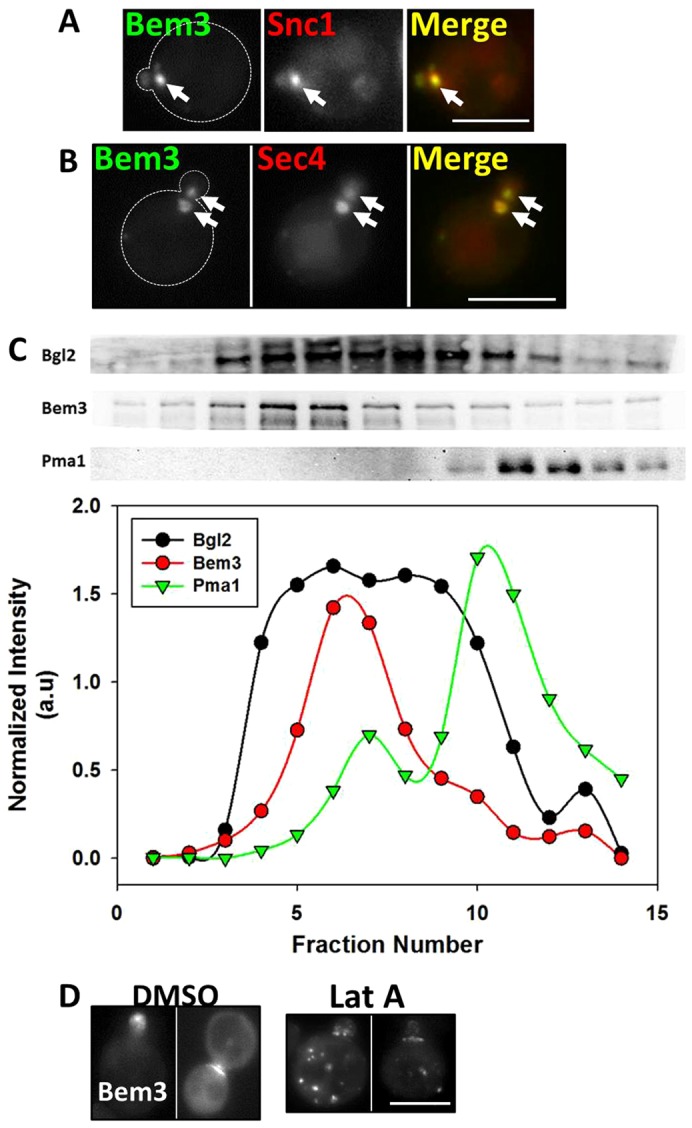
Fig. 4.

The Bem3-containing compartment colocalizes substantially with markers of the early endosome and secretory vesicles and traffics to sites of polarized growth on actin cables. (A) W303 cells transformed withYep13-GFP-Bem3 and pRS316-HA-mRFP-cSnc1were grown overnight in selective medium at 30°C and imaged at 100× using FITC and Rhodamine filters. Arrows point to the Bem3-containing compartment. (B) W303 cells expressing pGAL1.416-Bem3-GFP and pAD54-RFP-Sec4 were grown overnight in 2% glucose-containing medium at 30°C and transferred to 2% galactose medium for 2 hours before imaging at 100× using FITC and Rhodamine filters. Arrows point to the Bem3 compartment. Scale bar: 5 µm. (C) sec10-2 cells expressing HA-Bem3 from a GAL1 promoter were grown in glucose-containing medium overnight, then transferred to 2% galactose-containing medium and shifted to 37°C for 2 hours before cell lysis. The P3 pellet (see Materials and Methods) was subjected to Percoll density gradient fractionation. Fractionation profiles of Bem3, Bgl2 and PmaI were determined by western blotting (upper panels) with appropriate antibodies (see supplementary material Table S3), followed by band densitometry (lower panels). (D) W303cells expressing Yep13-GFP-Bem3 were grown overnight in selective medium, followed by addition of 200 µM Latrunculin A in DMSO, or vehicle alone, for 60 minutes and imaged at 100× using a FITC filter. Scale bar: 5 µm.
