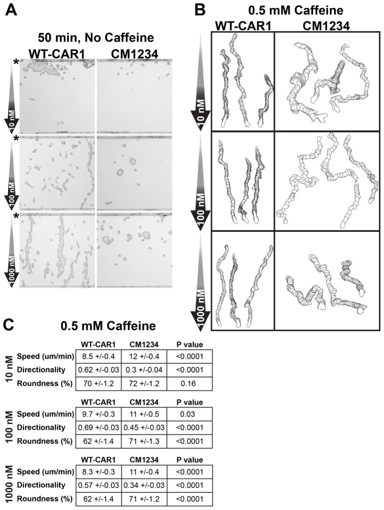
Fig. 1.

Loss of CAR1 phosphorylation impairs directional migration within a linear cAMP gradient. (A) Differentiated WT-CAR1 or CM1234 cells were exposed to linear cAMP gradients, as indicated, in an EZ-TAXIScan chamber and assayed over time for directional movement. Origin of cells in chamber is indicated (*). The 50 minute time point is shown. See supplementary material Fig. S2 for additional time points and supplementary material Movie 1 for an image sequence. (B) Differentiated WT-CAR1 or CM1234 cells were assayed as in Fig. 1A, but in the presence of 0.5 mM caffeine. cAMP concentrations used to initiate the gradient are indicated. See supplementary material Movie 2 for an image sequence. Shown are single-cell-outline traces using the DIAS software package. The cell tracings were stacked for 50 frames and compiled into one image to display morphometrics of the sequence. (C) Metrics were determined from tracings of ∼100 caffeine-treated cells in Fig. 1B using DIAS (see supplementary material Movie 2). Directionality (the net path length divided by the total path length) was measured using the chamber bottom as the reference point; a value of 1.0 corresponds to a completely straight path, whereas smaller values indicate a meandering path. 100% roundness indicates a circular cell, whereas 0% corresponds to a cell having no effective width or enclosed measurable area. P values of Student t-tests are shown.
