
Fig. 1.
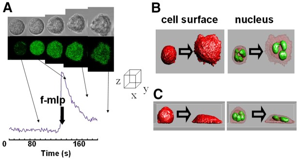
Rapid cell spreading in neutrophils is induced by a Ca2+ signal. (A) The upper series of images show the phase contrast images and the lower series the fluo4 intensity (as a readout of Ca2+ concentration) of an individual human neutrophil undergoing cell spreading in response to f-mlp (1 µM). The graph shows the time course of cytosolic free Ca2+ concentration with times at which the images were taken indicated and the time of addition of f-mlp shown. (B,C) The cell surface (stained with DiI) and the nucleus (stained with acridine orange) have been reconstructed from confocal optical slices of an adherent but not ‘spread’ and a spread cell and are shown in orthogonal planes (B,C) as isosurfaces (Imaris). Each of the examples shown are representative of at least 20 experiments using neutrophils taken from at least four different donors.