Fig. 4.
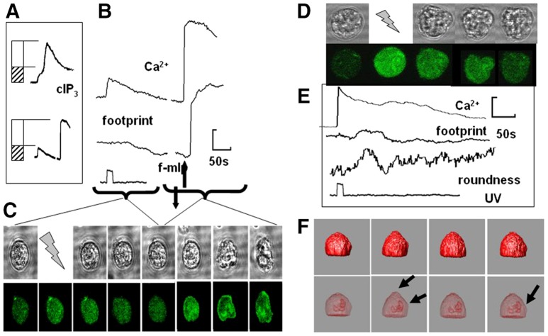
Factors affecting neutrophil spreading. The effect of uncaging Ca2+ (nitr5) to a level of the initial (release) phase of the caged IP3-induced Ca2+ signal. (A) The matching of initial (release) phase of the caged IP3-induced Ca2+ signal (upper graph ‘cIP3’) and nitr5 uncaging followed by addition of f-mlp (lower graph). (B) Graphs are showing the effect of f-mlp addition from A in more detail, with the accompanying effect on the cell ‘footprint’ and (C) images of the cell and its cytosolic free Ca2+ from which the data was taken. (D–F) The effect of the pre-treatment of neutrophils with cytochalasin B (5 µg/ml) before uncaging IP3: (D) a series of phase contrast and fluo4 images, (E) quantification of cytosolic free Ca2+, footprint and cell roundness [taken as (cell perimeter/π.max cell dimension)], and (F) changes in the reconstructed isosurface of the cell and the position of the nucleus, with the arrowheads indicating local sites of cell deformation. An example of the ‘wriggling response’ to IP3 uncaging in cytochalasin-B-treated neutrophils is shown in supplementary material Movie 2.

