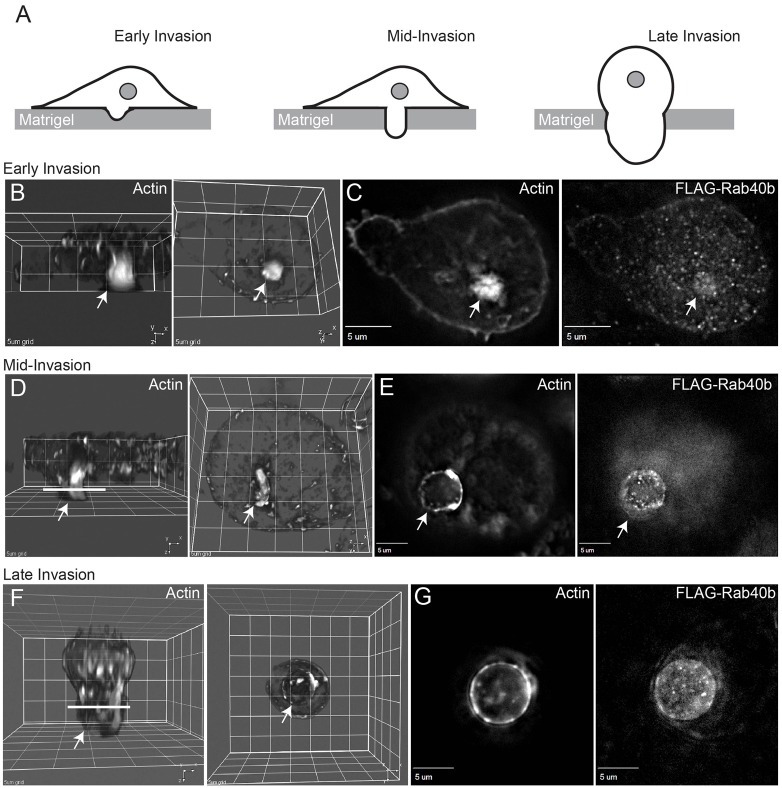
Fig. 6.
Localization of Rab40b-containing organelles during cell invasion in vitro. MDA-MB-231 cells stably expressing FLAG–Rab40b were seeded on Matrigel-coated filters containing 8 µm pores. Cells were incubated for either 24 hours (B,D) or 36 hours (F). Cells were then fixed and stained with Rhodamine-phalloidin or anti-FLAG antibodies. Drawings in A depict the invasion stages imaged in panels B,D,F. Arrows in all images indicate invadopodia or pseudopodia (probably derived from invadopodia). B,D,F are 3D rendering of images shown in C,E,G and show cells from Z-Y (left panels) and X-Y (right panels) planes. Lines in D,F indicate the level of the optical sections depicted in E,G.

